19世纪中叶,人类可能就已经见过线粒体。但碍于当时的固定和染色技术的局限,细胞生物学家们并没有明确的鉴别出细胞内的多种颗粒小体(cytologists)[1]。
一般认为,第一位对线粒体进行明确分离和描述的研究者是Kollike,他在1888年从昆虫飞行肌中提取出这种颗粒体。此后他还发现了这种小颗粒体的一些特性[2] [3]。
1898年,德国科学家Benda给这个小颗粒体起名线粒体(mitochondrion),尽管当时这个名字在同一批候选清单(肌质体sarcosome、原生粒bioblast、杆状物chondriocont等)中并不突出,但此后广为接受并沿用至今。它的词源是希腊语中的”线“(mitos)和”颗粒“(chondros),这个组合很好的体现了线粒体的形态多样性[4]。
1914年,Lewis MR和Lewis WH使用简单的光学显微镜,描述了细胞质内的线粒体大小、性状和位置的变化[5]。1934年,相差显微镜问世,刷新了对线粒体运动方面的认知。1941年,Michel首次展示了减数分裂过程中细胞内线粒体的快速运动。此后的二十多年间,很多研究沿用了相衬成像技术,但是这一技术不能完全明确识别出各种细胞器,观察线粒体的热情逐渐冷却。
荧光显微镜再次点燃对线粒体动态的观察。线粒体特异性荧光染料主要是亲脂性阳离子染料(如罗丹明123,Mito Tracker),这些染料虽然能够让细胞器在原位发出荧光,但是它们具有细胞毒性,并且很容易在强光条件下发生漂白。以绿色荧光蛋白(GFP)为基础的细胞成像技术改变了这一局面。1995年,Rizzutoet等首次使用GFP作为线粒体标记(mito-GFP),翻开了看见的全新一页。
1914年,Lewis和Lewis就已经观察到线粒体是一个多形性的(pleomorphic)细胞器。也就是说,一个给定细胞内的线粒体群体都是由一群形态各异且实时变化着的线粒体混合而成的。
但是,一个细胞中具体有多少个线粒体?它们是均匀的分散在细胞中还是缠绕粘连成一个整体呢(spatial arrangements)?这就需要引入两个新的词汇:一个细胞内所有线粒体的总称:线粒体系(chondriome)和它们的总体形态(gross morphology)。
在动物和酵母细胞中,线粒体通常是长管状的或者网状的[6],或呈一个连续矩阵(matrix continuity)[7]。
而在高等植物中,数百个甚至上千个球形或香肠状的线粒体离散分布在细胞内,形成一个不连续的整体(discontinuous whole),如下图中一个典型的拟南芥(Arabidopsis)叶肉细胞内离散着大约120个叶绿体和数百个线粒体[8]。
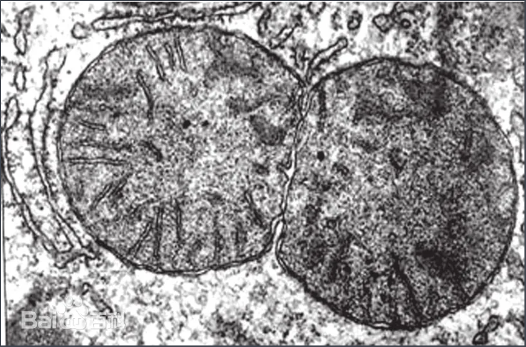
图1、正在分裂的线粒体

图2、0.2%健那绿染色10x100(目镜x物镜)观察的小鼠肺线粒体(荔园生物所示)
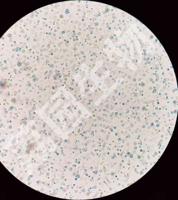
图3、0.2%健那绿染色10x100(目镜x物镜)观察的小鼠肝(冻存2d)线粒体(荔园生物所示)
^Logan, D.C., 2003. Mitochondrial dynamics. New Phytologist 160, 463–478. https://doi.org/10.1046/j.1469-8137.2003.00918.x
^Lehninger, A.L., 1964. The mitochondrion: molecular basis of structure and function. W. A. Benjamin, New York.
^Tribe, M.A., Whittaker, P.A., 1972. Chloroplasts and mitochondria: by Michael Tribe and Peter Whittaker, Institute of Biology’s studies in biology. Arnold, London.
^Faitg, J., Lacefield, C., Davey, T., White, K., Laws, R., Kosmidis, S., Reeve, A.K., Kandel, E.R., Vincent, A.E., Picard, M., 2021. 3D neuronal mitochondrial morphology in axons, dendrites, and somata of the aging mouse hippocampus. Cell Reports 36, 109509. https://doi.org/10.1016/j.celrep.2021.109509
^Lewis, M.R., Lewis, W.H., 1915. Mitochondria (and other cytoplasmic structures) in tissue cultures. American Journal of Anatomy 17, 339–401. https://doi.org/10.1002/aja.1000170304
^Logan, D.C., 2006. The mitochondrial compartment. Journal of Experimental Botany 57, 1225–1243. https://doi.org/10.1093/jxb/erj151
^Eisner, V., Cupo, R.R., Gao, E., Csordás, G., Slovinsky, W.S., Paillard, M., Cheng, L., Ibetti, J., Chen, S.R.W., Chuprun, J.K., Hoek, J.B., Koch, W.J., Hajnóczky, G., 2017. Mitochondrial fusion dynamics is robust in the heart and depends on calcium oscillations and contractile activity. Proceedings of the National Academy of Sciences 114, E859–E868. https://doi.org/10.1073/pnas.1617288114
^Logan, D.C., 2006. Plant mitochondrial dynamics. Biochimica et Biophysica Acta (BBA) - Molecular Cell Research, Mitochondrial Dynamics in Cell Life and Death 1763, 430–441. https://doi.org/10.1016/j.bbamcr.2006.01.003
^Chowdhary, S., Madan, S., Tomer, D., Mavrakis, M., Rikhy, R., 2020. Mitochondrial morphology and activity regulate furrow ingression and contractile ring dynamics in Drosophila cellularization. Mol Biol Cell 31, 2331–2347. https://doi.org/10.1091/mbc.E20-03-0177
^Jung Kim, M., Ho Kang, K., Kim, C.-H., Choi, S.-Y., 2008. Real-time imaging of mitochondria in transgenic zebrafish expressing mitochondrially targeted GFP. BioTechniques 45, 331–334. https://doi.org/10.2144/000112909